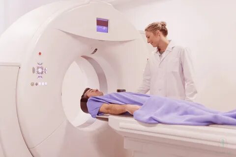
Computerized (or computed) tomography, also previously referred to as Computerized Axial Tomography (CAT) scan is an X-ray process that blends a variety of X-ray images using computer technology to produce cross-sectional images and, in the event of need three-dimensional views of internal organs and organs of the body. The Dr Ali Holistic provide the best priapus shot in USA. Computerized tomography is typically identified by its abbreviated name, CT scan or CAT scan. CT scans are used to identify abnormalities in the body. CT scan can be used to identify abnormal and normal tissues in the body, and/or aid in the treatment process by helping to ensure the correct placement of medical instruments or treatments.
A huge donut-shaped X-ray scanner or machine captures pictures of X-rays at various angles throughout the body. These images are processed computer software to create photographs of the body’s cross-section. In each of these images, the body is viewed like the”X-ray “slice” of the body that is then recorded on film. The recorded image is referred to as a tomogram. “Computerized axial tomography” refers to the tomogram that is recorded “sections” at different levels of the body.
Imagine your human body in the form of a loaf bread, and you’re looking at the one end of that loaf. When you cut off each bread slice you will be able to observe the entire surface of the slice, from the crust all the way to the middle. The body appears in CT scan slices in the same way beginning from the surface to the center of the body that is being studied. When these levels are “added” together, a 3D image of the organ or an abnormal body structure could be taken.
Which one is better, CT or MRI?
In general the CT as well as MRI scans are fairly secure. However, there could be issues. MRI scans are not conducted on patients who have aneurysm clip (clips of the vessels inside the brain) in the absence of being confirmed to be MRI secure, since the clips could be removed and the patient might suffer from bleeding in the brain.
Another issue associated with MRI can be the existence of certain defibrillators or cardiac pacemakers because magnetic fields can cause malfunctions within these battery-powered devices. Metal devices can be affected by a magnetic field such as that there are metal shavings inside an organ, eye, or even the extremity, could be removed due to magnets. Furthermore, other containers that are made of metal (like oxygen tanks) must be kept out of MRI machines as they could be drawn by the magnet and cause injury or death to the patient.
CT scans aren’t afflicted with the same issues, however they expose the patient to radiation, even though they’re a fairly small dose. Certain kinds of CT scans may not be suitable during pregnancy.
What’s the goal of the purpose of a CT scan?
CT scans are used to study inside the structures and internal organs of different areas of the body. The head is one of them which is where injuries from trauma (such such as blood clots, or skull fractures) as well as tumors and infections are identified. In the spine bone structure, the vertebrae is easily determined, as can the anatomy of intervertebral discs and the spinal cord. In actual fact, CT scan methods can be utilized to precisely measure the bone density when the evaluation of osteoporosis.
Sometimes it is the case that contrast substance (an an X-ray color) is added to the spinal fluid in order to improve the quality of the scan as well as the diverse structural relationships between the spinal cord, the spine and the nerves that it runs through. Contrast material can also be administered intravenously or via different routes prior to getting the CT scan (see below). CT scans can also be used in the chest to detect cysts, tumors or infections that are suspected by a chest Xray. Abdominal CT scans can be useful in determining the anatomy of the body, which includes imaging the gallbladder, the liver pancreas, spleen, kidneys, uterus, aorta and the ovaries. CT scans of this region can be used to confirm that there is no cancers, infections or abnormal anatomy changes to the body that are due to trauma.
It is completely painless and offers extremely precise images of body structures, in as well as guiding the radiologists through specific procedures, like biopsies for cancerous areas and the removal of body fluids for different examinations, as well as the drainage of abscesses, which are deep inside the body. A lot of these procedures aren’t invasive and have drastically reduced the need for surgeries to achieve the same objective.
Are there any risks involved in getting an CT scan?
The CT scan can be a low-risk procedure. The most frequent issue can be a reaction that is averse to contrast material that is intravenous. Contrast intravenous is typically an iodine-based fluid that is inserted into the vein. This causes numerous organs and structures like the kidneys or blood vessels, more evident in the CT scan. There could be an underlying itching and hives, or a rash or feeling of warmth across the body. They are typically self-limiting reactions that disappear quite quickly. In the event of a need, antihistamines could be prescribed to ease symptoms. An allergic reaction that is more severe to intravenous contrasts is known as an anaphylactic response. If this happens it can cause severe hives, and/or breathing difficulty. This is a rare reaction however; it could be life-threatening if not dealt with. The use of corticosteroids or antihistamines as well as epinephrine are able to reverse this reaction.
Kidney toxicity that may cause kidney failure is a uncommon complication that can result from the contrast agent intravenous utilized during CT scans. Patients with diabetes, those that are dehydrated and those who have already suffered from an impaired function of the kidneys are more susceptible to this reaction. The latest Intravenous contrast agents are being created for example, like Isovue and Isovue, which have virtually removed this problem.
Radiation that a patient receives in the CT scan is low. In both non-pregnant and pregnant women, the scan hasn’t been demonstrated to cause negative results. When a female is expecting it could pose an increased risk for the fetus in particular during the first trimester the pregnancy. In the event that a person is expecting and is not aware of it, she must inform her doctor about her pregnancy and talk about different imaging options like ultrasound scan, which is not dangerous to the embryo. However, exposure to radiation during an CT scan can cause a small increase in the lifetime likelihood for developing cancer. The Dr Ali Holistic provide the best orgasmic shot in USA. The issue is generally regarded as more significant for children because the risk of cancer for radiation doses is greater for children than adults, and young patients live longer duration. But, the risk of radiation exposure must be evaluated against the benefits of making use of CT scanning to identify or treat illnesses. CT scanners can be altered to give exposures better suited to children. The majority of doctors recommend that radiation exposure for patients be reduced to a minimum. patients who “doctor shop” or repeatedly visit emergency rooms for an “CT” put themselves at risk of having radiation-related issues.
